You are under about 60 and have arthritis in only one half of the knee — usually the inner side.
Your legs are bow-shaped or knock-kneed, and the alignment is concentrating your weight onto the worn compartment.
You want to stay physically active in a way that a knee replacement would limit — manual work, running, farming, sport.
You are too young for a replacement to be a sensible one-off, and would otherwise face a revision later in life.
The cartilage on the other side of your knee is still healthy.
It is probably not right if…
Arthritis affects the whole knee rather than one compartment.
The knee is stiff, or cannot fully straighten.
You smoke and are not willing to stop — smoking substantially reduces the chance of the bone healing.
You are unable to manage six to eight weeks on crutches.
You are over about 65 with widespread wear, where a replacement is the better and more predictable operation.
The idea in one paragraph
Most knee arthritis is not evenly spread. In the majority of people the inner half wears out first, because the shape of the leg sends more load through it. If your legs are slightly bow-shaped, that effect is amplified: you are walking through the worn side of your knee thousands of times a day. An osteotomy changes the angle of the bone so that the load transfers to the healthy side. The worn cartilage is not repaired — it is unloaded, and an unloaded joint hurts far less.
The two operations
High tibial osteotomy (HTO)
For bow legs with inner-compartment wear — by far the more common. A precise cut is made in the top of the shin bone, the angle corrected, and the bone held with a plate and screws while it heals. Load moves from the worn inner side to the healthy outer side.
Distal femoral osteotomy (DFO)
For knock knees with outer-compartment wear. The same principle applied to the lower end of the thigh bone, shifting load from the outer side back towards the inner.
Both are planned from full-length standing X-rays, on which the mechanical axis of the whole limb is measured and the exact correction calculated in degrees before you come to theatre. During the operation the correction is confirmed with live imaging.
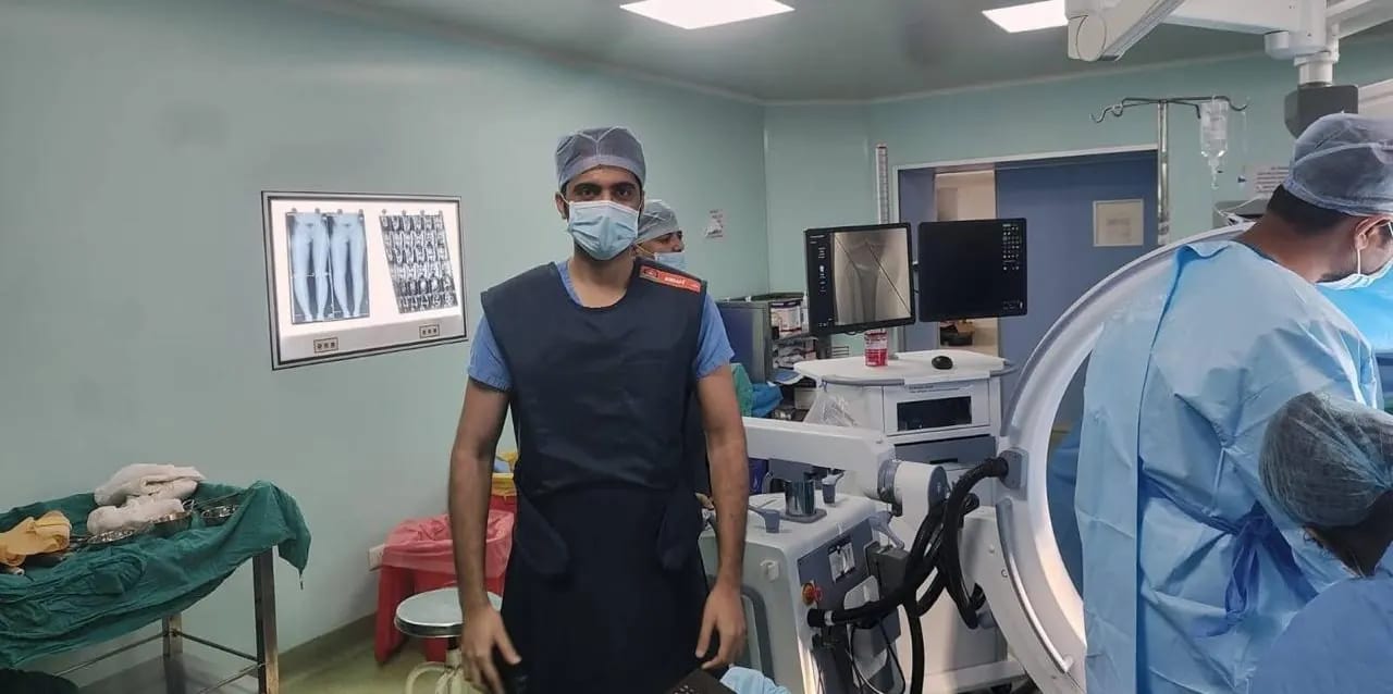
Live imaging in theatre. Alignment is planned on standing X-rays beforehand and confirmed in degrees during the operation — this is a millimetre-accurate procedure, not a judgement by eye.
Osteotomy or replacement?
Aspect
Osteotomy
Partial or total replacement
Typical age
Under 60
Over 60, or lower demand
Arthritis pattern
One compartment only
One or all compartments
Your own joint
Kept
Resurfaced or replaced
Running and impact
Allowed
Generally discouraged
Heavy manual work
Allowed
Often limited
Recovery
6–9 months
3–4 months
Crutches
6–8 weeks
1–3 weeks
Typical lifespan
10–15 years, often longer
15–25 years
What comes next
A replacement remains possible
Revision replacement
Both are good operations. They suit very different people.
What else counts as joint preservation
Osteotomy is the largest of a group of operations whose shared purpose is keeping the joint you were born with, rather than replacing it.
Meniscus repair — preserving the shock absorber instead of removing it.
Cartilage restoration — microfracture or grafting techniques to resurface a defined defect.
Patellofemoral realignment — correcting a kneecap that tracks badly and is wearing unevenly.
Ligament reconstruction — an unstable knee damages its own cartilage with every episode of giving way.
Loose body removal and treatment of a cartilage flap before it does further harm.
Risks
Delayed healing or non-union of the bone cut, which is uncommon but much more likely in smokers.
Over- or under-correction of the alignment, which is why the planning is done on standing films and checked in theatre.
Infection, and the possibility of needing the plate removed.
A blood clot in the leg.
Irritation from the plate, which some patients choose to have removed after the bone has healed.
Nerve irritation causing temporary numbness or weakness in the foot, usually recovering.
Continued progression of arthritis over the following years, since the joint has been unloaded rather than made new.
What recovery actually looks like
Written as a plan, not a promise. Your own timeline will be adjusted to what is found during the operation and how you progress — but this is the shape of it.
Day 0–3
In hospital
Two to three nights. You will be up with a physiotherapist on day one, learning to walk with crutches while protecting the leg. Pain is controlled with a combination of medications.
Standing and walking with crutches on day one
Partial weight-bearing technique taught
Knee movement started immediately
Home by day two or three
Week 1–6
Protecting the bone
Crutches with a defined amount of weight through the leg while the bone cut heals. Movement is encouraged throughout — it is the load that is restricted, not the bending.
Full range of knee movement
Back to desk work at around three to four weeks
Wound healed and reviewed
X-ray at six weeks to check healing
Week 6–12
Back onto the leg
Once the X-ray shows the bone uniting, weight-bearing progresses to full and crutches are discarded. Strengthening begins properly. Most patients drive from eight to ten weeks.
Off crutches
Walking normally
Driving again
Static cycling and pool work
Month 3–6
Rebuilding
The bone continues to consolidate and the muscles rebuild. Walking distance grows steadily, and most people notice the pain relief clearly by now.
Long walks without pain
Return to physical work
Full gym-based strength programme
Stairs comfortable in both directions
Month 6–9
Full activity
By six to nine months the bone is fully healed and there are no restrictions. Unlike after a replacement, running and impact sport are permitted.
Running permitted
Return to sport
No activity restrictions
Plate removal considered if it is causing irritation
Patients who had this
Told he needed a knee replacement at 47. He kept his own knee.
“Two doctors had told me a replacement was the only option, and that I would have to give up the farm work. Here I was shown standing X-rays and told the wear was only on the inner side, and that my bow legs were the reason. The eight weeks on crutches were long. But I am back on the land and I still have my own knee.”
Bhavesh D., 47 · Farmer
Bardoli · 22 months after treatment
Outcome. High tibial osteotomy correcting the alignment away from the worn inner compartment. Off crutches at eight weeks, back to full agricultural work by seven months, no restriction on physical labour.
A consultation means your imaging on the screen, a plain explanation of what is actually wrong, and an honest opinion on whether you need surgery — including when the answer is no.